|
One of the most serious diseases affecting adults, especially caucasians, asians and females, is osteoporosis, or literally 'holes in the bone'. Osteoporosis is a loss in bone mass leading to thin, fragile bones especially in the spine and the femur. Estrogen loss, inactivity, vitamin D deficiency, lowered calcium and protein intake, and smoking are all factors leading to this disease. With this disease, the number of bone fractures dramatically increases as pathological fractures, (spontaneous breaks without apparent injury) begin to occur.
| Differences Between Normal and Osteoporotic Bone |
|
|
Rickets is a disease that occurs in children and is marked by the inability of bones to calcify, or harden. The bones then soften and the weight-bearing bones of the legs begin to bow. Rickets is due to a lack of calcium or vitamin D, which aids in the absorbtion of calcium into the blood stream. It is not seen in the USA very often due to a wide avalability of vitamin D fortified foods and dairy products, but remains a problem in third world countries.
| X-Ray of Child with Rickets |
|
|
Joint Disorders:
Osteoarthritis: degeneration of the joints, usually weight-bearing ones; common in elderly people
Bursitis: inflamation of bursa sacs within the joints, usually caused by excessive stess; creates pain, redness, swelling, and stiffness around the affected joint
Rheumatoid Arthritis: most serious form arthritis, extremely painfull, recurring inflammation can lead to deformaties; is an autoimmune disorder affecting the synovial membranes.
Tuberculosis and Syphilis can also create an infection within bones (osteomyelitis) which are difficult to treat and may cause skeletal difformaties.
| Affects of Rheumatoid Arthritis on a Joint |
|
|
Osteosarcoma - Bone Cancer:
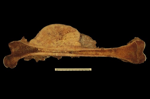
Osteosarcoma usually occurs in the femur or around the knee and the tibia, or around the shoulder. The typical patient is between 20-25 and the symptoms include deep, ill-defined pain and swelling in the area of the tumor. Sometimes the patient will have a pathologic fracture (see osteoporosis). An x-ray typically shows the tumor and a CAT scan is used to look for metastases, or to see if the cancer is spreading. A biopsy is then performed and the treatment plan is created. Chemotherapy is common and surgery to remove the tumorous materials is regular. Statistics shows that 60-70% of patients with non-metastatic osteosarcomas live without a recurrence of the disease. If the patient lives more than 5 years after diagnosis they have a very high chance of never relapsing.
| X-Ray of Femur & Skull with Bone Tumors |
|
|
| Femur with Osteosarcoma |

|
|

